You may have dental X-rays at a new patient appointment. UK radiation rules require each exposure to be clinically justified. The dentist first builds a picture from your concerns, dental and medical history, examination and any useful recent records.
If an image is likely to influence diagnosis or treatment, the dentist can explain what it is intended to show. If it would add little, it may be deferred. This individual approach avoids both unnecessary exposure and the risk of missing information that cannot be seen directly.
A mirror and good lighting reveal exposed tooth surfaces, gums and other mouth tissues. They cannot show every area between teeth, beneath a restoration, inside a tooth or within the jaw. A radiograph can add information when the expected clinical benefit justifies the small radiation exposure.
Possible reasons include checking for decay between teeth, investigating pain, assessing bone levels around teeth, looking at roots, planning some extractions or restorations, and reviewing development or unerupted teeth. The image selected should match the question. A small intra-oral view and a panoramic image are not interchangeable.
Dental X-rays also have limits. Early cracks, bite problems and many soft-tissue conditions may not appear. A dark or light area needs interpretation alongside the examination; it is not a diagnosis by itself.
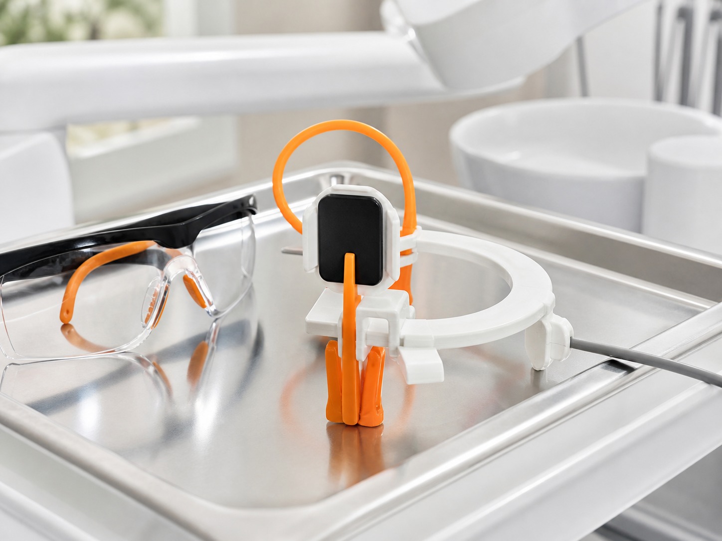
Bitewing images show the crowns of upper and lower back teeth together and can help assess decay between teeth and crestal bone levels. Periapical images show a tooth from crown to root and surrounding bone, which can be useful for pain, root treatment or local infection.
A panoramic radiograph provides a broad two-dimensional view of the jaws and teeth. It may help with unerupted teeth, broad assessment or some surgical planning, but it is not the best image for every small area. CBCT produces three-dimensional information and generally involves more exposure than routine two-dimensional dental views, so it needs a specific indication.
The dentist should use the lowest exposure consistent with obtaining diagnostic information. Equipment checks, staff training, positioning and selecting the appropriate field all contribute to optimisation.
For an intra-oral image, a small sensor or plate is positioned inside the mouth while the X-ray tube is aligned from outside. You will be asked to keep still for a brief exposure. Tell the operator if a strong gag reflex, limited opening, pain or another accessibility need makes positioning difficult; alternative techniques or adjustments may be possible.
For a panoramic image, you usually stand or sit while the machine moves around your head. Jewellery, glasses or removable appliances may need to be taken out because they can obscure the image. The operator positions your head carefully and asks you to remain still.
The result is checked for diagnostic quality. Repeating an image is not routine: it should happen only when the first image cannot answer the justified question and the benefit of another exposure remains greater than the risk.
Tell the dental team if you are or may be pregnant, particularly when asked under the practice procedure. UKHSA patient guidance describes the risk from dental X-rays during pregnancy as negligible to minimal, but every exposure still needs justification and optimisation. The dentist can discuss the benefit of imaging now, any appropriate alternative and whether delay would affect your care.
When arranging a new-patient visit, tell the practice if another dentist has recent images. With your permission, records may be requested. The date, image type, diagnostic quality and reason for the current assessment determine whether they remain useful.
An old image can provide valuable comparison even when a new view is justified. Conversely, a recent image may not show the area now causing symptoms. Avoid assuming that transferring records guarantees no further imaging or that a full set must be repeated.
Keep details of previous implant, root canal, gum or orthodontic care. A concise timeline can help the clinician interpret both the images and the present examination more accurately.
X-rays use ionising radiation, so the exposure is not described as zero-risk. The practical safeguard is to take an image only where the likely health benefit outweighs the radiation detriment and to keep exposure as low as reasonably practicable for the clinical purpose.
The exact dose varies by image, equipment, settings and patient. Comparing a dental image with background radiation can be a useful explanation, but it should not become a blanket justification. Ask what the proposed image is for, whether recent images exist and how the result could affect care.
The UK Health Security Agency provides accessible information on medical imaging benefits and risks. Your dentist can explain the recommendation in the context of your mouth rather than relying on a generic dose table.
A comprehensive first visit normally includes your priorities, medical history, previous dental experience and an examination of the teeth, gums, bite and soft tissues. Screening findings and risk assessment shape discussion of prevention, urgent needs and options. Images are one evidence source within that process.
The exact sequence depends on symptoms. Acute pain may need focused diagnosis before a full long-term plan. Complex treatment may require further records or a separate planning visit. Read about a new patient examination in Kingston to understand the appointment rather than treating X-rays as the purpose of the visit.
After assessment, the dentist should explain significant findings and next steps in understandable terms. You can ask to see the images and ask how they support the proposed plan.
No. The decision is based on individual clinical need, examination findings, risk and useful existing records.
You can discuss and decline a proposed exposure. Ask about its purpose and implications; without necessary information, the dentist may be unable to diagnose or provide certain treatment safely.
UKHSA describes dental X-ray risk in pregnancy as negligible to minimal. Tell the team so the exposure can be considered under the practice procedure and justified for your circumstances.
There is no single interval for everyone. Decay risk, age, examination findings and previous images influence timing.
No. It complements the history and examination; some cracks, bite problems and soft-tissue conditions may not be visible.
Thames Street Dental can review your concerns and available records, examine your mouth and explain whether any imaging is clinically justified.







 020 8546 6546
